
Figure 6.
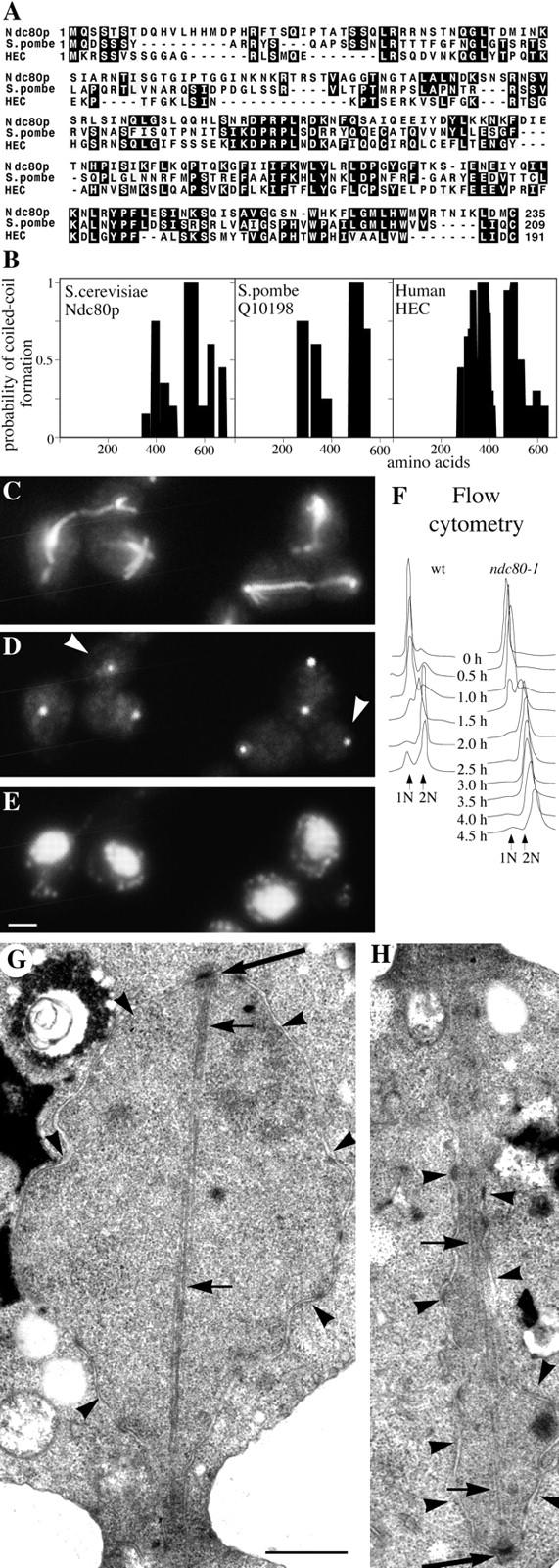
Characterization of Ndc80p. (A) Homology between the NH2-terminal domains of Ndc80p, S. pombe Q10198, and human HEC; (B), comparison of the coiled-coil domains (determined by Paircoil) of the same three proteins; (C–E), immunofluorescent staining of ndc80-1 after 2 h at 36°C with anti-tubulin (C); anti-Tub4p to stain the SPBs (D); and DAPI to stain DNA (E). (D) White arrowheads, SPBs not associated with chromosomes; (F) flow cytometry to measure DNA replication in wild-type and ndc10-1 cells synchronized by elutriation and released at 36°C; (G and H), electron micrographs of serial thin sections from the same large budded ndc80-1 cell synchronized by α-factor and released at 36°C for 2.5 h. G shows one end of a postanaphase spindle and H the other end of the same spindle in the adjacent section. Large arrows, SPBs; small arrows, microtubules; arrowheads, nuclear pores and nuclear membrane. Bars: (C–E) 2 μm; (G and H) 0.5 μm.