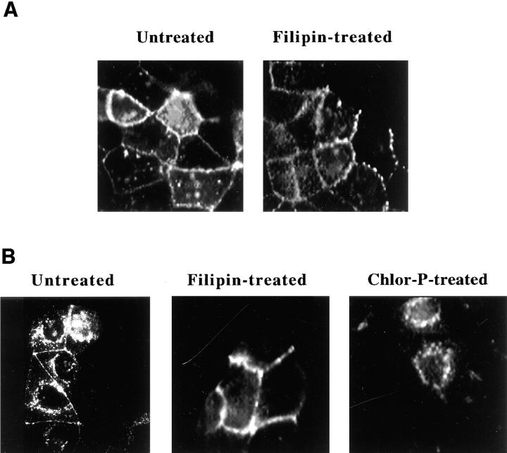
Figure 3.
Effects of filipin and chlorpromazine on the distribution of rhodamine-conjugated CT-B in CaCo-2 cells detected by direct fluorescence microscopy. Cells were incubated for 1 h at 37°C in MEM, Hepes, and 0.01% BSA with no addition; 1 μg/ml filipin; or 25 μg/ml chlorpromazine. The cells were then labeled with 5 nM Rh-CT-B for 30 min at 15°C in fresh medium in the presence or absence of the same effectors. Cells were washed once and incubated an additional 30 min at 4°C (A) or 37°C (B) in fresh medium with effectors and fixed. The distribution of Rh-CT-B was then visualized by direct fluorescence as described in Materials and Methods.

