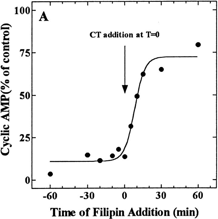
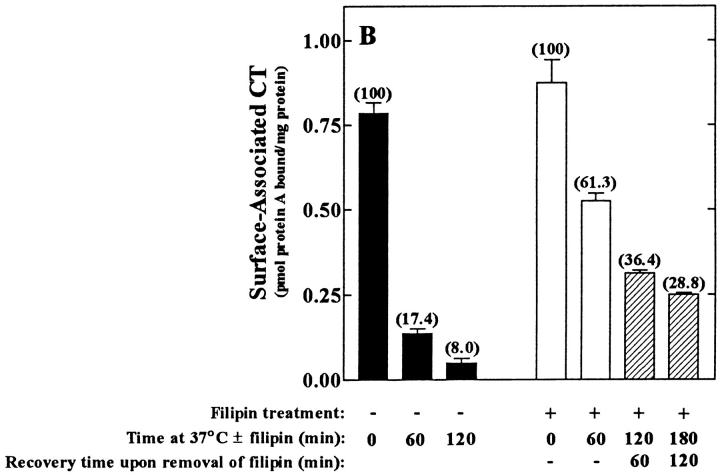
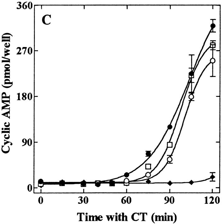
Figure 7.
Filipin-mediated inhibition of CT activity in CaCo-2 cells is both rapid (A) and reversible (B and C). (A) Filipin (1 μg/ml) was added to cells either before (− values) or after (+ values) the addition of CT (0.03 nM, final). All the cells were exposed to the toxin for 2 h at 37°C and assayed for the accumulation of cAMP. Results are expressed as a percentage of cAMP formed in the absence of filipin. (B) Cells were treated in the absence (filled bars) or presence (open and cross-hatched bars) of 1 μg/ml filipin for the indicated times at 37°C, chilled to 4°C, and incubated with 10 nM CT for 1 h ± filipin. Cells were either exposed to anti-CT-A1 at 4°C (zero time) or incubated at 37°C with the same additions for the indicated times before antibody exposure. Some of the filipin-treated cells (hatched bars) were washed and incubated in filipin-free medium for an additional 60 and 120 min at 37°C before the addition of antibody at 4°C. Immunodetection of surface-bound CT was quantified with 125I-protein A as described in Materials and Methods. (C) Cells were treated in the presence (♦, ○, □) or absence (•) of 1 μg/ml filipin for 1 h at 37°C and some of the cells were either washed briefly (○); or, for 30 min (□) in filipin-free medium at 37°C. Then all of the cells were incubated with 0.03 nM CT for the indicated times at 37°C and assayed for cAMP accumulation as described in Materials and Methods.


