
Figure 2.
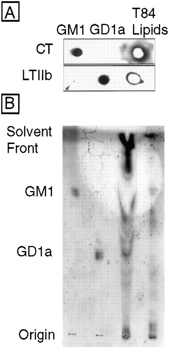
Ganglioside- enriched lipid extracts of T84 cell monolayers contain both GM1 and GD1a. (A) Lipid extracts prepared from 1,500 cm2 confluent T84 cells (third lane) and purified GM1 (first lane) and GD1a (second lane) standards (300 ng each) were applied to activated silica gel plates, processed as described in Materials and Methods, and ligand blotted with CT (1 nM, top) or LTIIb (1 nM, bottom). Toxin binding to ganglioside extracts was assessed by application of specific antitoxin antibodies, followed by HRP-conjugated secondary antibody, and development using enhanced chemiluminescence. (B) Size resolution of T84 cell gangliosides by thin layer chromatography and resorcinol spray. GM1 standard (first lane), GD1a standard (second lane), T84 cell (4,500 cm2) lipid extract lower phase Folch cut (third lane), and upper phase Folch cut (fourth lane). In this preparation, the bulk of acidic glycolipids including GD1a remained associated with the lower lipid soluble phase because of conditions of Folch extraction (low salt and pH). Gangliosides migrating with GD1a are apparent in the third lane (and faintly in the fourth). Gangliosides migrating with GM1 are apparent in the fourth lane.