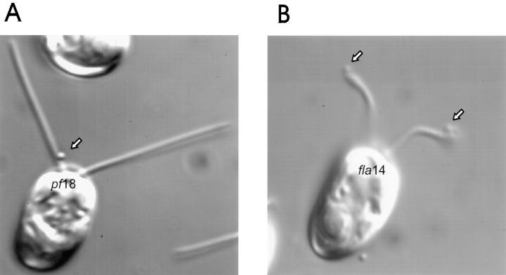
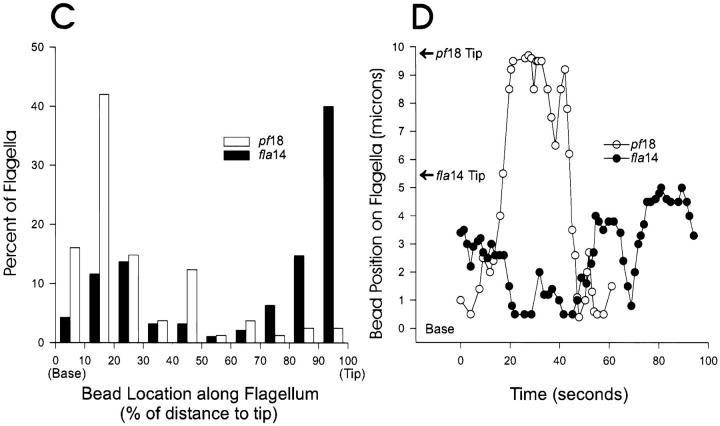
Figure 7.
Beads move in both directions but accumulate at the tips of fla14 mutant flagella and at the base of pf18 flagella. Polystyrene beads (Polysciences, Warrington, PA, 0.3 μm in diameter) were added to pf18 and fla14 (V64) cells. In general, beads (arrows) tended to be located at the bases of pf18 flagella (A) but at the distal tips of fla14 flagella (B). (C) Bead position was quantitated by continuously recording while moving through microscope fields and then measuring the position of bound beads with respect to the ends of the flagella. As soon as a flagellum with attached bead came into view, the position of the bead on the flagellum was measured; only one measurement was made per flagellum. On average, pf18 flagella were 9.8-μm long and fla14 flagella were 4.9-μm long. To compare data from different length flagella, the bead position was converted to percent of length and plotted as a histogram. (D) Beads bound to flagella of pf18 (open circles) and fla14 (closed circles) are actively translocated in both directions. Moving beads were observed by DIC microscopy and videotaped. In each case, the position of the bead on the flagellum was measured and plotted as a function of time. Note that beads bound to both pf18 and fla14 flagella make long, smooth runs in both directions. The pf18 sequence was atypical in that the bead was at the flagellar tip during much of the recording time.


