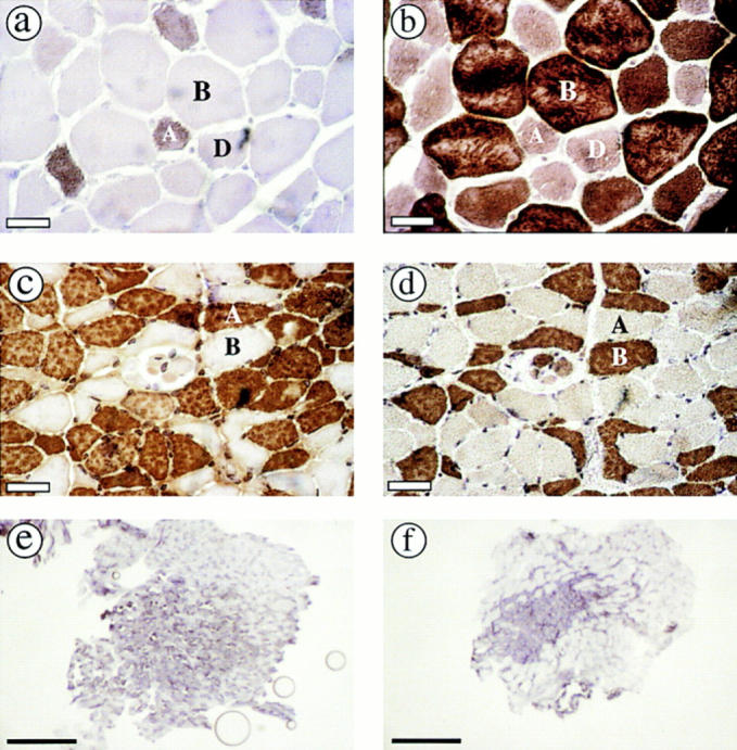
Figure 3.

MyHC isoform content of psoas muscles from wild-type and MyHC-IId null mice determined by immunohistochemistry. Serial sections of psoas muscle from wild-type (a and b) and MyHC-IId null (c and d) mice (8-wk-old males) were stained with antibodies to MyHC-IIa (a and c) or MyHC-IIb (b and d). Positive fibers were visualized by peroxidase staining. Sections of psoas muscle from wild-type (e) and MyHC-IId null (f) mice were stained by NADH-TR and photographed at low magnification (4×). Bars: (a–d) 30 μm; (e and f) 0.5 mm.
