
Figure 7.
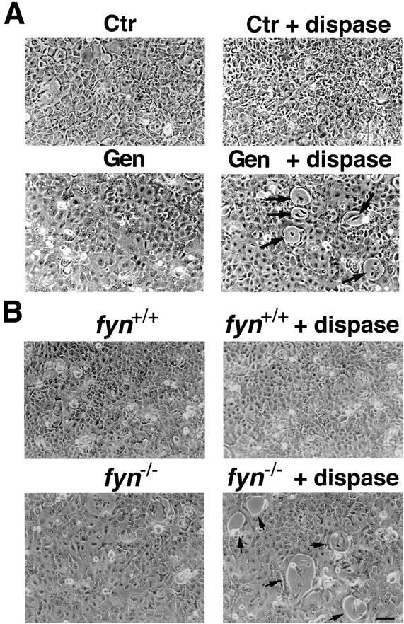
Decreased strength of calcium-induced cell adhesion as a consequence of tyrosine kinase inhibition or lack of the Fyn kinase. (A) Primary keratinocytes under growing conditions were either tested as untreated controls (Ctr) or pretreated for 2 h with Genistein. Incubation was then continued for additional 24 h under high calcium conditions. Cultures were examined as such (left panels) or after dispase treatment for 5 min (right panels). Arrows, the focal areas of cell detachment that occurred in the tyrosine kinase inhibitor-treated cultures. No such areas were evident in control cultures even after prolonged dispase exposure (>30 min). Instead, control cells eventually detached from the dish as a confluent sheet. Similar results were observed with keratinocyte cultures switched to high calcium conditions for only 9 h (not shown). (B) Primary keratinocytes derived from fyn−/− mice and wild-type littermates were exposed to high calcium concentrations (2 mM) for 9 h. Cultures were examined as such (left panels) or after treatment with dispase for 5 min (right panels). Arrows, the focal areas of cell detachment that occurred in the fyn−/− cultures already at this time. There were no cells missing in the monolayer of fyn knockout keratinocytes before dispase treatment. As we previously reported (Calautti et al., 1995), the fyn knockout keratinocytes fail to stratify and are larger than normal, which explains the different morphological appearance of these cultures relative to the wild-type controls even before dispase treatment. Similar results were observed in two other independent experiments. Bar, 60 μm.