
Figure 3.
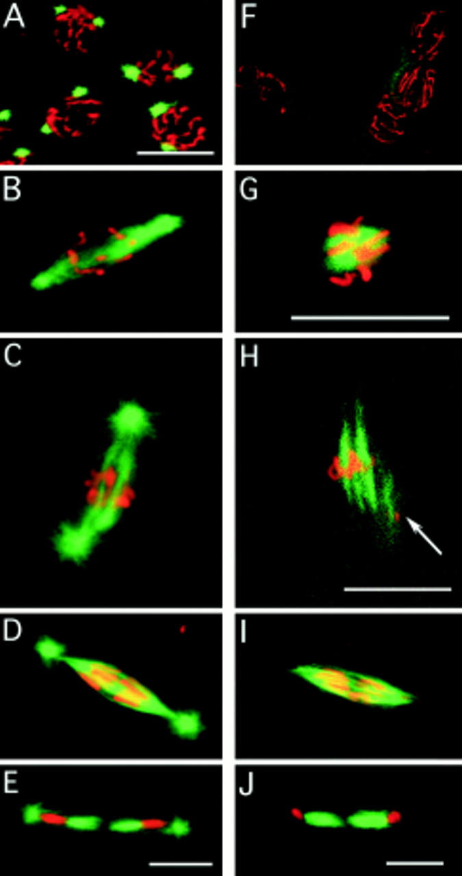
Spindle assembly in the absence of centrosomes. Embryos are double stained for their DNA (red) and microtubules (green). Fertilized embryos: (A) In prophase, the chromosomes are condensed on the nuclear membrane, and the centrosomes have migrated to approximately a 180 degree position. (B) After nuclear membrane breakdown, the centrosomes of fertilized embryos nucleate microtubules that are preferentially directed toward the chromosomes. (C) Metaphase fertilized embryos have a spindle between the two centrosomes. (D) In early anaphase, the spindle is sharply pointed, and the chromosomes are starting to segregate. (E) In telophase, the midbody (central spindle) has formed between the two daughter nuclei of the division. Unfertilized embryos: (F) In prophase, the chromosomes are condensed on the nuclear membrane, but there are no MTOCs. (G) Tubulin is apparent on and near the chromosomes as the spindle starts to assemble. (H) Arrays of microtubules arise from the chromatin. The arrow marks a second metaphase nucleus partly in the plane of focus with a spindle also assembling. The close apposition of nuclei is typical of unfertilized embryos. (I) The appearance of the anaphase spindle is very similar to the anaphase spindle of fertilized embryos, except that centrosomes with astral microtubules are conspicuously absent. (J) In telophase, the midbody (central spindle) has formed between the two daughter nuclei of the division, even though there are no centrosomes. Bars, 10 μm.