Figure 6.
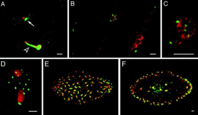
Free MTOCs are present in fertilized Sciara embryos. These embryos are double stained for their DNA (red) and microtubules (green). (A) During telophase of female meiosis II, the centrosome associated with the decondensing male pronucleus is nucleating a large aster (arrow), and a large midbody separates the female pronucleus and the second polar body. (B) The asters of eight MTOCs are in the plane of focus surrounding the fusing pronuclei. Three polar bodies reside at the cortex. (C) Magnified view of pronuclei. (D) During interphase of nuclear cycle 2, the two nuclei are encompassed by 16 MTOCs. E and F depict surface and medial views of a nuclear cycle 8 embryo and demonstrate that the free MTOCs do not migrate to the cortex. Bars, 10 μm.

