Figure 7.
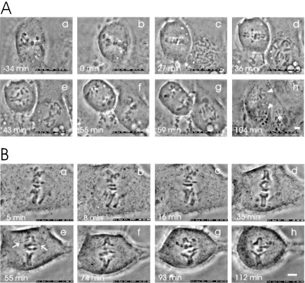
Microinjection of mitotic PtK1 cells with anti-p55CDC antibodies impairs normal progression of late mitotic events. (A) Slow separation of sister chromatids and impeded exit from mitosis by anti-p55CDC microinjection. The cell was microinjected with anti-p55CDC antibodies (1.5 mg/ ml in the micropipette) at early prometaphase (6 min after NEB). (a) The first image was taken 13 min later when the cell was at late prometaphase. (b) The chromosomes reached the metaphase plate. (c) Anaphase initiated after a short delay of ∼26 min. (c–g) Separation of sister chromatids was very slow, requiring over 50 min before the sister chromatids reached the spindle poles. The cell failed to undergo cytokinesis, and exited mitosis as a binucleate cell (arrowheads in h). To the right of the microinjected cell, an untreated cell progressed through a normal mitosis. The asterisk in h denotes one of the progeny of this normal division. (B) Inhibition of sister chromatid separation by anti-p55CDC microinjection. The cell was injected at late prometaphase with anti-p55CDC antibodies (1.5 mg/ml). (a) The first evidence of chromatid segregation was seen 5 min after the start of metaphase (time point = 0 min). (b–h) Despite pulling forces evident from stretching of the kinetochore regions (e, arrows), full separation of sister chromatid arms was never achieved. Bars, 5 μm.

