Figure 9.

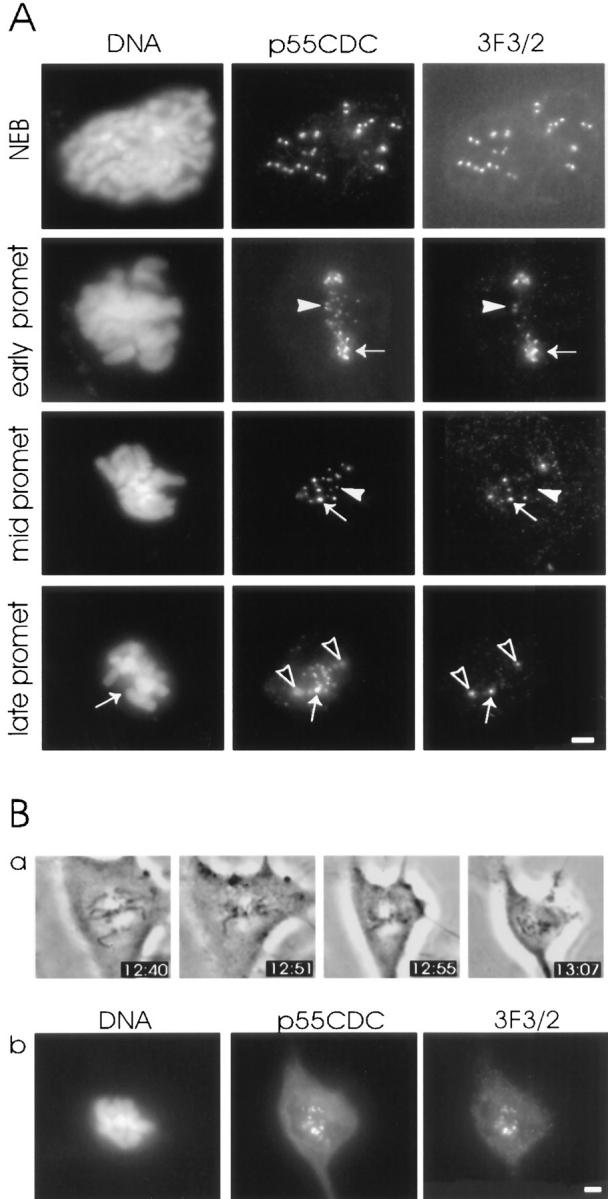
Expression of 3F3/2 phosphoepitope at the kinetochores of anti-p55CDC microinjected PtK1 cells. (A) Similar to p55CDC signal, the intensity of the fluorescent label of 3F3/2 epitope varies among chromosomes with different positions along the spindle. The kinetochores of chromosomes closer to spindle poles possess brighter 3F3/2 and p55CDC signals (arrows in early prometaphase and mid prometaphase rows) compared with kinetochores of aligned chromosomes (arrowheads in early prometaphase and mid prometaphase rows). At late prometaphase and metaphase the 3F3/2 signal disappears from the kinetochores of aligned chromosomes, while p55CDC is still present at all kinetochores. One misaligned chromosome having a bright p55CDC signal also shows strong labeling with the 3F3/2 antibody (arrows in late prometaphase row). The spindle poles that are labeled by both p55CDC and 3F3/2 are denoted with open arrowheads. (B) The 3F3/2 phosphoepitope expression reappears at the kinetochores of anti-p55CDC injected cells at metaphase if the spindle is destroyed with nocodazole. (a) At 12:35 the early prometaphase cell was injected with antibodies against p55CDC (1.0 mg/ml). At 12:55, all the chromosomes were aligned at the spindle equator. Nocodazole (5 μg/ml) was added at 12:57. (b) The cell was fixed 15 min later, and was immunolabeled to detect injected anti-p55CDC and the 3F3/2 phosphoepitope. Bars, 5 μm.
