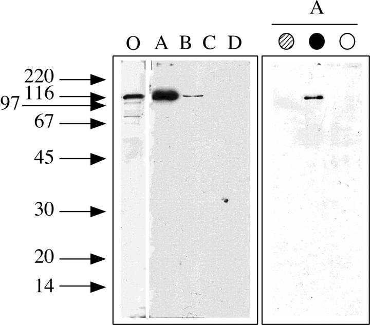
Figure 2.
Western blot analysis of the 104-kD protein in the nuclear extract separated by phosphocellulose chromatography and in the proteins coimmunoprecipitated with the estrogen receptor in the flowthrough fraction. Arrows on the left side indicate the migration of standard proteins of the indicated molecular weight (kD). (Left) SDS-PAGE followed by Western blot analysis with mAb 1032 to the major vault protein of MCF-7 cells nuclear extract (lane O) and of the flowthrough (lane A), 0.3 M (lane B), 0.5 M (lane C), or 0.75 M KCl (lane D) fractions of the phosphocellulose chromatography of nuclear extract. (Right) SDS-PAGE followed by Western blot analysis with mAb 1032 to the major vault protein of proteins immunoprecipitated with the mAbs to estrogen receptor AER314 (gray circle) and AER317 (solid circle) or control IgG (open circle) of the flowthrough fraction of the phosphocellulose chromatography of nuclear extract.

