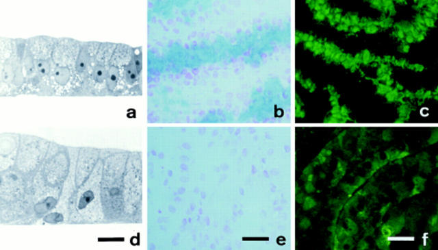
Figure 2.
Effects of GalNAc-α-O-benzyl on cell morphology and mucus expression in HT29-RevMTX10−6 mucus-secreting cells. Cells were cultured in the absence or under permanent exposure to 2 mM GalNAc-α-O-benzyl and then analyzed after 21 d in culture. Left column, light microscopy of thin sections of the cell layer perpendicular to the surface of the flask. Middle column, alcian blue staining of cryostat sections of cell layers from the same cultures, counterstained with nuclear red. Right column, indirect immunofluorescence staining with pAb L56/C of cryostat sections of the same cell layers. (a–c) Untreated cells; (d–f), cells treated with GalNAc-α-O-benzyl. Bars: left column (17 μm); middle and right columns (40 μm).

