
Figure 4.
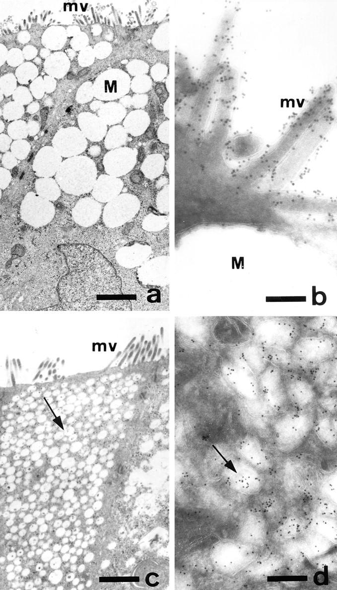
Ultrastructural morphology and localization of DPP-IV in control and GalNAc-α-O-benzyl–treated HT-29 mucus-secreting cells. HT29-RevMTX10−6 cells were analyzed after 21 d of culture in the absence (a and b) or presence (c and d) of 2 mM GalNAc-α-O-benzyl. (a and c) Standard transmission electron microscopy of sections perpendicular to the bottom of the flask showing the apical microvilli in both control and treated cells, the accumulation of mucus droplets (M) in the apical compartment of control cells, and the very numerous vesicles (arrow) in treated cells. (b and d) Immunogold labeling of DPP-IV using mAb HBB 3/775/42: in control cells the gold particles are strictly restricted to the microvilli and absent from the mucus droplets; in treated cells the gold particles are associated with the cytoplasmic vesicles. Similar results were obtained with MUC1 and CEA (data not shown). Bars: (a and c) 1.2 μm; (b) 0.16 μm; (c) 0.28 μm.