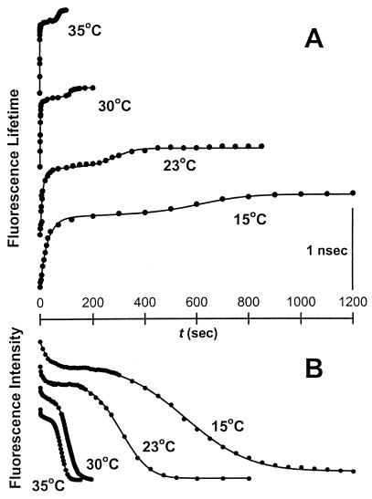
Figure 2.
Time courses of lipid and proton transfer during PEG-mediated vesicle fusion at different temperatures. PEG and vesicles were mixed at time zero. Lipid mixing (A) and proton transfer (B) were monitored by an increase in DPHpPC fluorescence lifetime and a decrease in HPTS fluorescence intensity, respectively (6). The initial lipid mixing/proton transfer indicates outer leaflet mixing and initial pore formation caused by the formation of intermediate I1. The delay suggests closing of initial pores and a transition to more stable intermediate I2. A slow lipid mixing and proton transfer during the delay was discussed in terms of lipid mass movement from outer to inner leaflet and occasional transient pore formation through the septum (see Discussion). Formation of FP causes the final step of lipid mixing/proton transfer: inner leaflet mixing, and complete proton transfer. The origins of each curve have been displaced to display data obtained at all four temperatures on the same plot. Each data set was fit to the kinetic model described in Materials and Methods, and the fitted curves are shown as solid lines. The activation energies (see Fig. 3) and rate constants at 35°C are listed for each step in Table 1.

