
Figure 2.
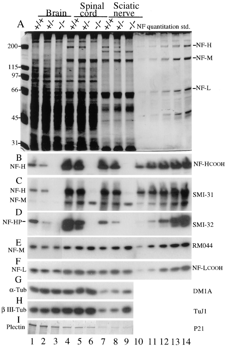
Levels of neurofilament subunits NF-L, NF-M, and NF-H in mice with zero, one, or two copies of a disrupted NF-H gene. (A) Total tissue extracts from 5-wk-old brain, spinal cord, and sciatic nerves were fractionated on 7% SDS–polyacrylamide gels and stained with (A) Coomassie blue or (B–I) electroblotted to nitrocellulose. (B) NF-H detected with a peptide antibody recognizing the extreme COOH terminus of NF-H (Xu et al., 1993); (C) phosphorylated NF-H and NF-M detected with monoclonal antibody SMI-31; (D) nonphosphorylated NF-H detected with monoclonal antibody SMI-32; (E) NF-M detected with monoclonal antibody RM 044 (Tu et al., 1995); (F) NF-L detected with a polyclonal peptide antibody recognizing the COOH terminus of NF-L (Xu et al., 1993); (G) α-tubulin detected with monoclonal antibody DM1A; (H) the neuron-specific class III, β-tubulin isotype with mAb TuJ1 (Lee et al., 1990); and (I) plectin detected with polyclonal antiserum P21 (Wiche and Baker, 1982). (Plectin migrates with a mobility of ∼500 kD in brain and spinal cord but at ∼160 kD in nerve samples using both this antibody and monoclonal antibody 10F6 [Foisner et al., 1991]; not shown.) Lanes 10–14, quantitation standards for the neurofilament subunits provided by a twofold dilution series of a neurofilament preparation. Molecular masses (kD) are indicated at left. (Lanes 1–3 of D–F represent four times longer exposures than lanes 4–14.)