Figure 4.
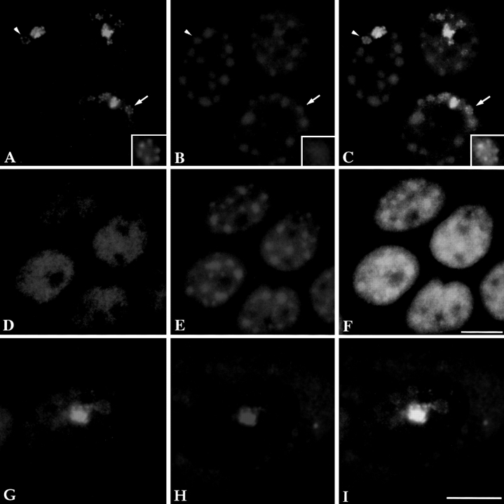
The structure of the PNC changes upon transcription inhibition by α-amanitin (an inhibitor of RNA polymerase II and/or III) in cells transiently expressing GFP– PTB (A). When double labeled with the Sm antibody (B), the extended PNC appears to form rosettes surrounding the round Sm clusters (C, inset). This change was not observed in cells that do not contain a PNC (D–F), suggesting that the change is specific to a reorganization of the PNC. When the distribution of GFP–PTB (G) and CUG-BP/hNab50 (H) was compared upon transcription inhibition by α-amanitin, a similar reorganization of the PNC in the nucleus was observed for both hnRNP proteins (G–I) in spite of the fact that the majority of the CUG-BP/hNab50 protein becomes predominantly localized in the cytoplasm (H). Bar, 10 μm.

