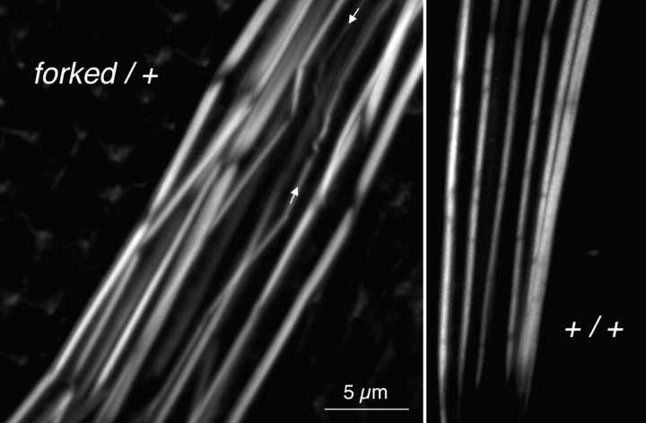
Figure 4.
Confocal images of portions of two bristles stained with fluorescent phalloidin to indicate the actin distribution. Left, forked36a/+ expressing less than the normal amount of forked proteins. Of interest is that the bundles are swollen relative to the wild type (right, same magnification). Some of the modules composing the bundles in the heterozygote are present at an acute angle, and some thin bundles are wavy in their trajectory (arrows).

