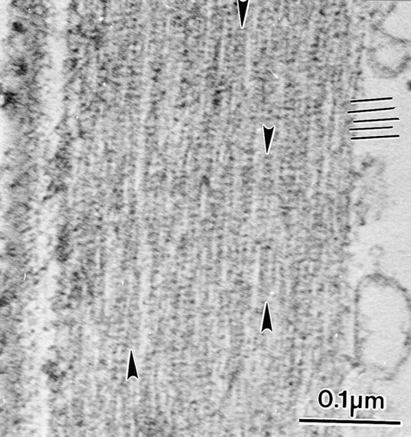
Figure 6.

Longitudinal section through an actin bundle in a bristle from the same mutants depicted in Figs. 4 and 5 where a reduced amount of forked protein is present. By turning the micrograph 90° one can see the 12-nm period due to fascin (black lines). A careful examination of this micrograph, however, reveals that the filaments are in small clusters (arrows). Thus, the 12-nm stripe extends across these clusters but does not extend across the space between adjacent clusters. The 12-nm stripe continues down the length of each cluster.
