
Figure 5.
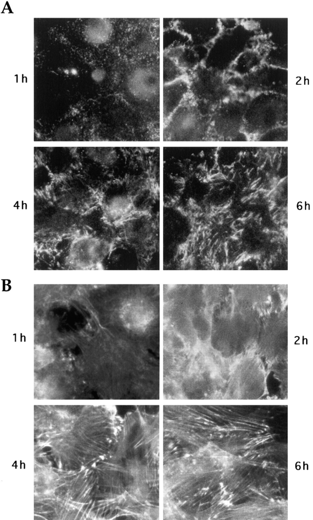
Time course of fibronectin matrix deposition and stress fiber organization after attachment of cells on substrate. Trypsinized KD fibroblasts were plated on a mixture of collagen IV and vitronectin in serum-free medium and stained after 1, 2, 4, or 6 h for cell surface fibronectin (A) or actin cytoskeleton (B).