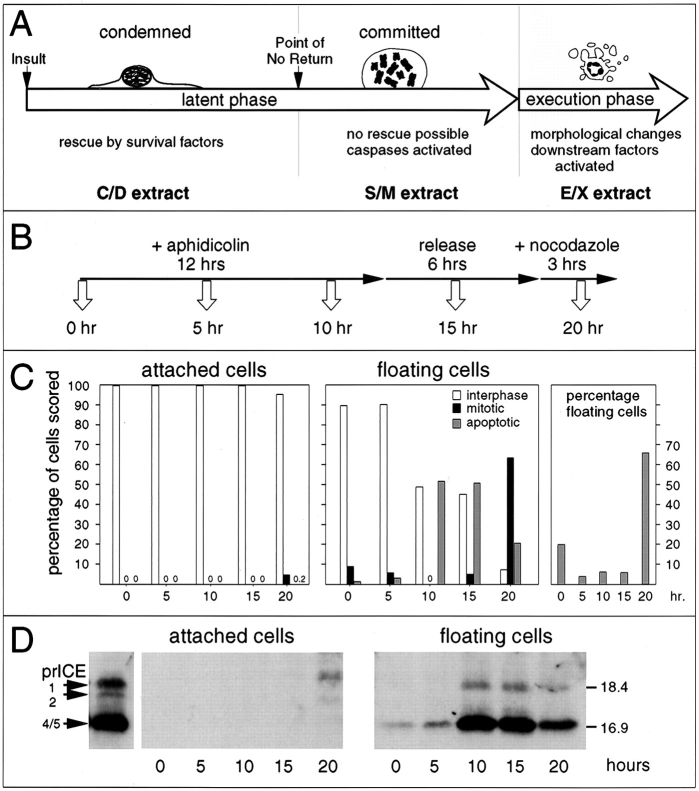
Figure 1.
(A) Diagram of apoptosis as a three stage process, with the latent phase being subdivided into condemned and committed stages. (B) The protocol used for harvesting of samples for examination of caspase activation. (C) The cells harvested at each time point were scored for their nuclear morphology, based on DAPI staining. (D) Caspase activity in whole cell lysates prepared from cells harvested at various times points after the addition of aphidicolin to the culture (time in hours shown at bottom). Left, attached cells; right, floating cells. The left-most lane shows the profile of active caspases in S/M extract.

