Figure 2.
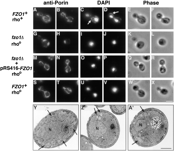
fzo1Δ cells exhibit defective mitochondrial morphology and lose their mtDNA. (A–X) Indirect immunofluorescence images of yeast cells stained with anti-porin antiserum to visualize mitochondrial membranes (leftmost columns), and DAPI to visualize nuclei and mtDNA (middle columns; white arrows in C and D indicate mtDNA nucleoids). Phase-contrast images of the same cells are shown in the rightmost columns. (A–F) Normal mitochondrial morphology and distribution of mtDNA nucleoids in a wild-type rho+ strain (JSY1812). (G–L) Mutant fzo1Δ rhoo cells (JSY1810) contained spherical or slightly elongated mitochondrial structures that lack DAPI-stained mtDNA. (M–R) Wild-type mitochondrial morphology, but not mtDNA, was restored in an fzo1Δ rhoo mutant strain (JSY2579) upon reintroduction of the wild-type FZO1 gene. (S–X) Mitochondrial morphology is normal in a wild-type rhoo strain (JSY2555) that lacks mtDNA. (Y–A′) Transmission electron micrographs of wild-type and fzo1Δ cells. (Y) Mitochondrial profiles in wild-type cells (JSY1812) are dispersed equally throughout the cell cortex and display numerous cristae (black arrows). (Z and A′) Mitochondrial profiles in fzo1Δ cells (JSY1810) are clustered together near the cell periphery and contain fewer cristae. N, nuclei; V, vacuoles; rho +, containing mtDNA; rhoo, lacking mtDNA. Bars: (A–X) 5 μm; (Y–A′) 1μm.

