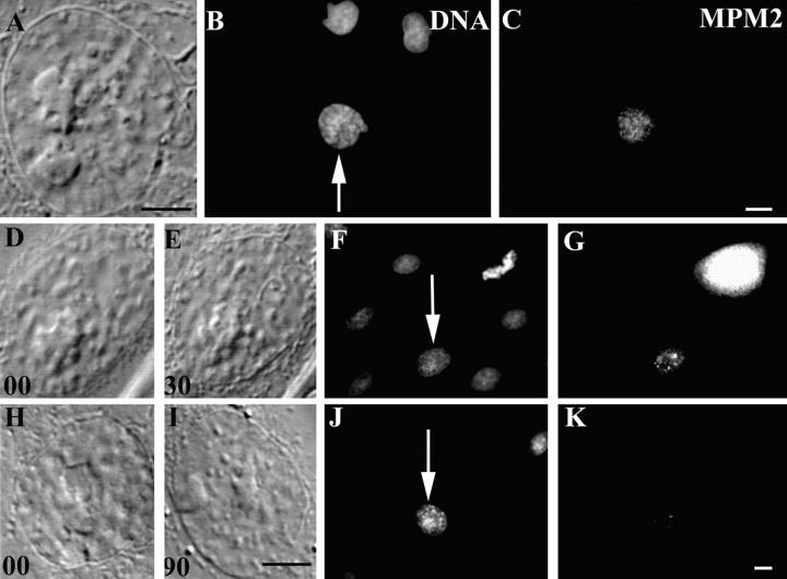
Figure 7.
The mitosis-associated phosphorylated epitopes recognized by the MPM2 antibody disappear from prophase nuclei as they return to interphase in response to nuclear irradiations. Conditions were the same as in Fig. 5 except that these cells were stained with a monoclonal antibody to MPM2 antigens. Early prophase (A and B, arrow) PtK1 nuclei contain appreciably more MPM2 epitopes than adjacent interphase nuclei (C). By contrast, similar prophase nuclei fixed 30 min after a nuclear irradiation (D–F) contain significantly fewer MPM2 epitopes (G), whereas those fixed after 90 min (H–J) largely lack these epitopes (K). As evident from the Hoechst image (F), the bright MPM2-positive cell in G is in metaphase of mitosis. Bars, 10 μm.

