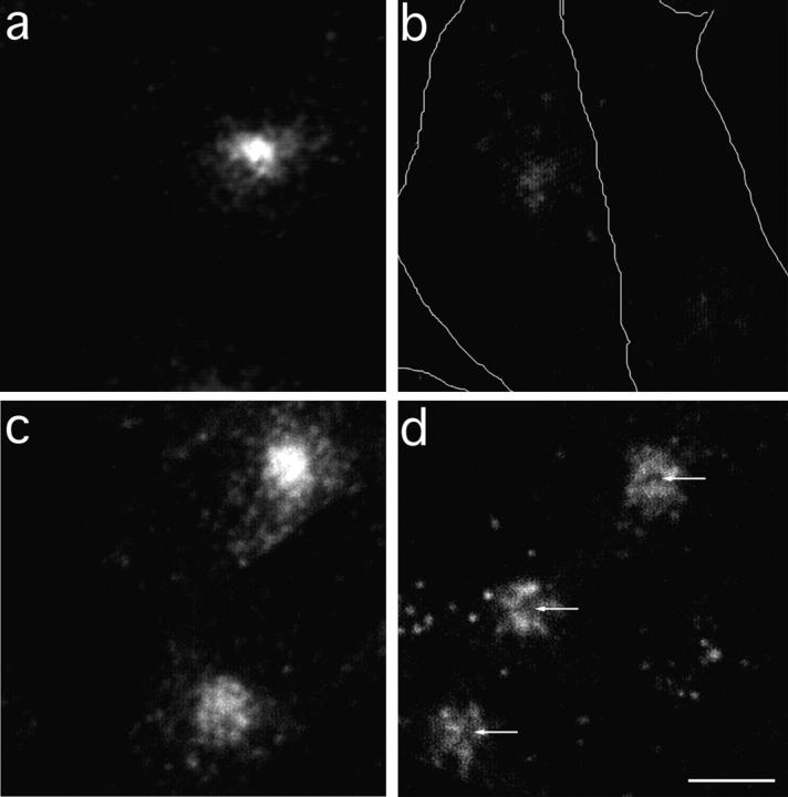
Figure 5.
TacTGN38 overlaps with Tf in the ERC, as shown by HRP ablation. Cells were incubated with 5 μg/ml Cy3-Tf for 30 min, and were then chased for 10 min with 50 μg/ml HRP-Tf with (a) or without (b) excess (5 mg/ml) unlabeled Tf. The cells were then treated with hydrogen peroxide and DAB. The reaction products of the DAB reaction quenched fluorescence from molecules contained in the same compartment as the HRP-Tf (b), and the unlabeled Tf competed with HRP-Tf for binding to the TR and prevented quenching of the Cy3-Tf fluorescence (a). The cell boundaries, determined by DIC microscopy, are shown in b. Cells were also incubated with 1 μg/ml Cy3 anti-Tac IgG for 10 min, and were then chased for 10 min with 50 μg/ml HRP-Tf with (c) or without (d) 5 mg/ml unlabeled Tf. The DAB reaction with the HRP-Tf in the ERC quenched the Cy3-IgG fluorescence in this compartment (d), leaving a dark area (arrows) surrounded by the ring staining of the Cy3-IgG characteristic of the TGN. The fluorescence images show a summation projection of slices in a 3-D stack obtained by confocal microscopy. Bar, 10 μm.

