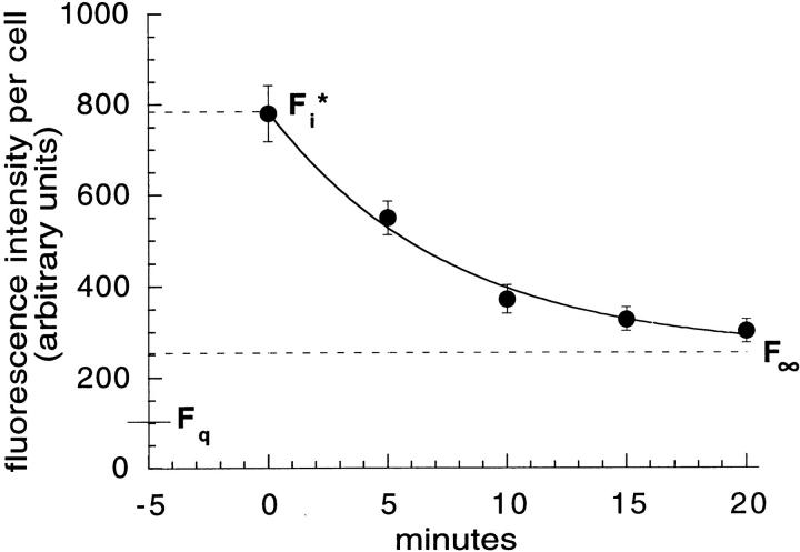
Figure 7.
18% of TacTGN38 is delivered to TGN with each round of endocytosis. Cells were labeled with 1 μg/ml F-anti-Tac IgG on ice, and were then warmed for 5 min, washed, and chased with 10 μg/ml anti-fluorescein antibodies. Fluorescence images were obtained by wide-field microscopy, quantified, and fit to a monoexponential decay. Data from a representative experiment are shown. The asymptote gave the residual fluorescence F∞, and a cell that was fixed and imaged without warming gave the initial cell-associated fluorescence, F i*. F q is the residual fluorescence after quenching surface bound anti-Tac. Error bars represent SEM.

