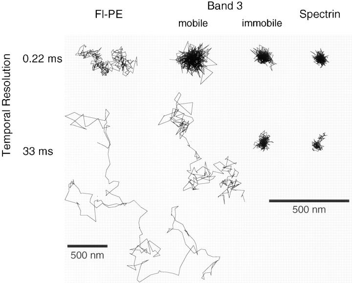
Figure 3.
Typical trajectories of band 3 that is (or is not) undergoing macroscopic diffusion, spectrin, and Fl-PE (artificially incorporated lipid) in erythrocyte membranes with time resolutions of 33 and 0.22 ms (total observation times of 6.7 s and 67 ms), respectively. The macroscopically mobile band 3 was undergoing apparent simple Brownian diffusion at a time resolution of 33 ms. However, the diffusion rate was slow compared with that of Fl-PE in the same time scale. Macroscopically immobile band 3 showed oscillatory movements at both time scales, which were similar to that of spectrin. Note that the magnification for Fl-PE with a 33 ms resolution is reduced by a factor of two. Bars, 500 nm.

