
Figure 9.
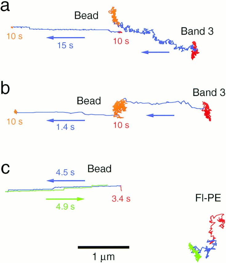
Examination of the viscous drag induced by the membrane skeleton dragging. (a and b) Latex bead attached to the membrane skeleton (Bead) was dragged using optical tweezers at a velocity of 0.15 μm/s (a) and 1.8 μm/s (b) for a distance of 2.5 μm as shown in the blue lines. Gold particles bound to band 3 that was undergoing macroscopic diffusion (Gold) followed the bead even at a slow rate of 0.15 μm/s. The movement of band 3 was like a superposition of hop diffusion and directed motion of the membrane skeletal network. (c) Effect of dragging the membrane skeleton on the lateral diffusion of lipid. When the latex bead attached to the membrane skeleton (Bead) was dragged at a velocity of 0.6 μm/s, gold particles attached to Fl-PE (Gold) did not exhibit any forced displacement (blue and green lines). Bar, 1 μm.