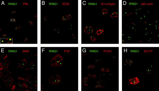
Figure 2.
The mammalian PcG complex forms a novel nuclear structure. The mammalian PcG complex was immunolocalized in human cells using the RING1-specific polyclonal antibody ASA3, and was stained using an FITC-conjugated secondary antibody. By double immunofluorescence microscopy, the staining pattern obtained for RING1 in 2C4 human fibrosarcoma cells was compared with that of known nuclear structures. The green signal shows labeled RING1, and the red signal given by Texas red–conjugated secondary antibodies shows immunolocalization of the following: (A) PML nuclear bodies from the anti-PML antibody 5E10 (Stuurman et al., 1992); (B) speckled domains recognized by the anti-SC35 antibody (Fu and Maniatis, 1990); (C) the snRNP family of proteins, recognized by the anti-Sm antigen antibody Y12 (Lerner et al., 1981); (D) coiled bodies recognized by the anti-p80 coilin antibody p-δ; (E) gems recognized by the anti-SMN antibody 2B1 (Liu and Dreyfuss, 1996); (F) PTB protein (Huang et al., 1997); (G) DNA replication foci detected using antibodies to PCNA; (H) sites of RNA synthesis localized with antibodies to in vivo–incorporated bromo-UTP nucleotides (note this panel is shown for U-2 OS cells). All images are a digital overlay of the two optical channels obtained from a single optical confocal section.

