
Figure 9.
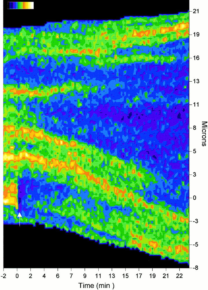
Mobility of E-cadherin puncta within the cell–cell contact interface. Fig. 9 shows a TIP scan of an entire contact during a photobleach-recovery experiment. A newly developing plaque in a 1.5-h-old contact was photobleached with a 2.8-μm-diameter bleach circle (0 mins, 0 μm) on the TIP scan (arrow). Images were collected every 16 s for 24 min at 0.11 μm/pixel. The fluorescence intensity scale bar ranges from 0–255 units divided into 15 colors.