
Figure 5.
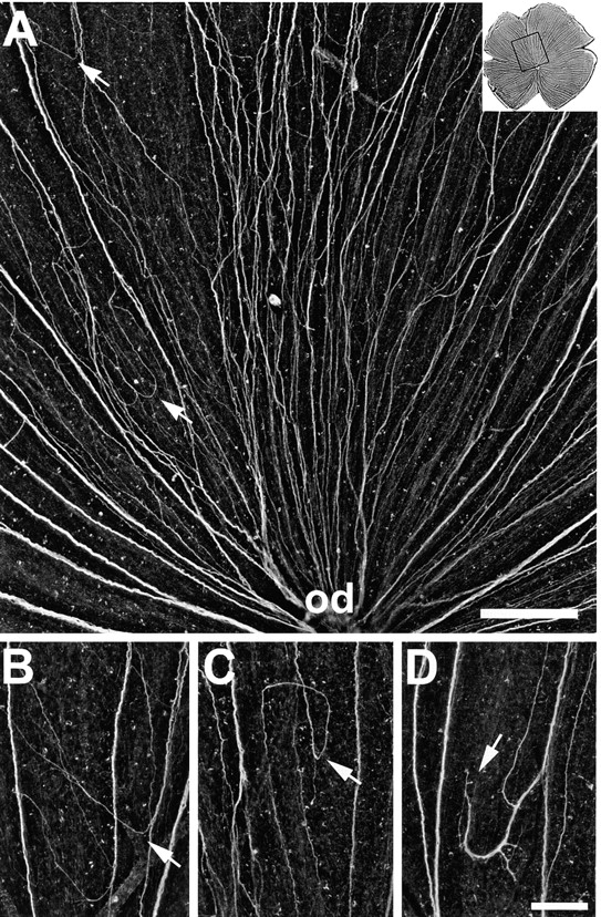
Aberrant routes in retinae injected with mAb N518 against Ig domain 2. (A) Higher magnification of the dorsal retina shown in Fig. 4, as indicated by the box in the upper right corner. Instead of growing directly towards the optic disk (od), young RGC axons form new subfascicles, change direction (arrows), and follow irregular pathways. (B–D) Examples of pathfinding errors. Young RGC axons depart from disk-oriented fascicles (arrow in B), grow back towards the retinal margin, establish circular routes (arrow in C), and form subfascicles that end in between fascicles (arrow in D). The retinal margin is up and the optic disk down. Bars, A, 200 μm; B–D, 100 μm.