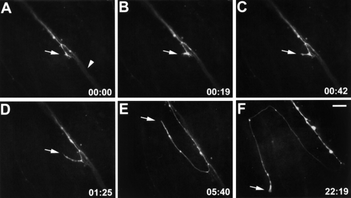
Figure 8.
Time-lapse sequence of an axon after mAb N518 injections. A DiO-labeled growth cone (arrow in A) elongates towards the optic disk (to the lower right) in association with other labeled axons of the same fascicle (arrowhead in A). This growth cone left its fascicle (arrow in B), turned away from the optic disk (C–E), turned again, and continued to grow outside the fascicle (arrow in F). Elapsed time is given in hours and minutes in the lower right corner. Bar, 20 μm.

