
Figure 1.
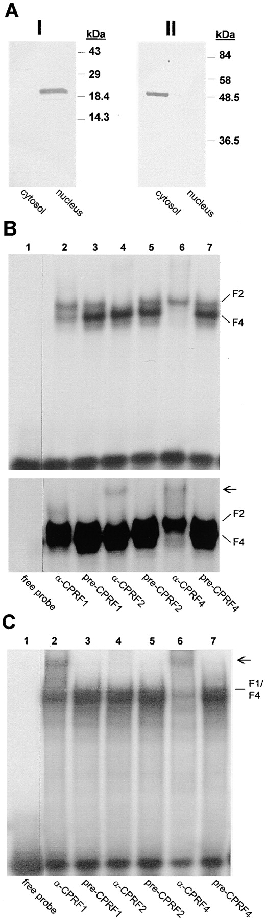
Analysis of the cytoplasmic and nuclear distribution of G-box–binding activities in dark-kept evacuolated parsley protoplasts. (A) Western blot analysis of cytoplasmic (cytosol) and nuclear (nucleus) extracts probed with histone 2A/2B (panel I) and tubulin (panel II) antibodies. In (I) 25 μg of protein per lane and in (II) 10 μg of protein per lane were loaded. In B and C autoradiograms of EMSSAs with 50 μg per lane of cytoplasmic and 20 μg per lane of nuclear extract are shown. For CPRF/antiserum interaction test the extracts were incubated for 10 min on ice with 1 μl of serum and the radioactive-labeled G-box probe before loading the samples on the gel (A and B, lanes 2–7). In B a 12- (top) and a 24-h (bottom) exposure of the identical shift gel is shown. In lanes 1, the binding reaction mix contained neither a serum nor protein (free probe). CPRF-containing DNA–protein complexes are marked (F1, CPRF1; F2, CPRF2; F4, CPRF4). Arrow, positions of supershifted DNA–CPRF complex.