
Figure 4.
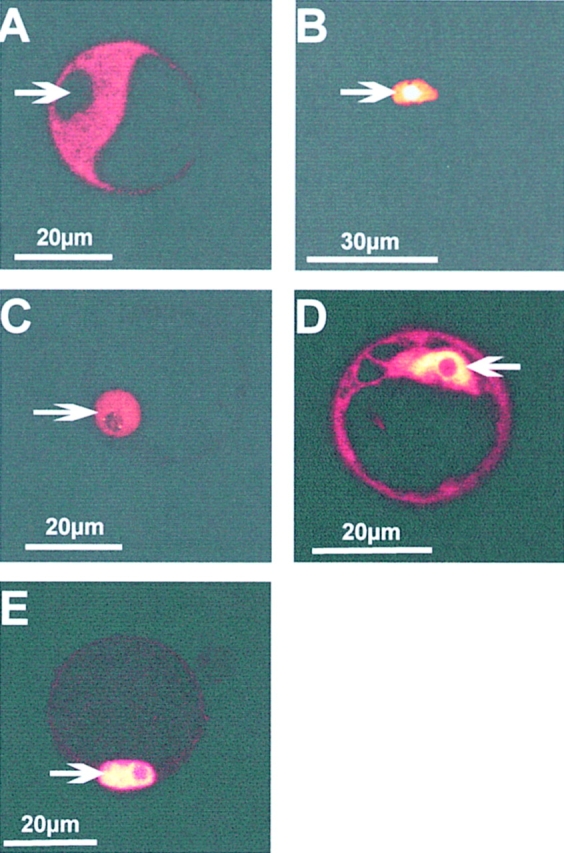
Intracellular sorting of GFP fusion proteins within parsley protoplasts. Confocal sections of parsley protoplasts transiently transformed with fusion constructs expressing phyA–GFP (A), NLS–GFP (B), CPRF1–GFP (C), and CPRF2–GFP (D and E). After transformation by electroporation protoplasts were kept for 16 h in darkness (A–D) or continuously irradiated with UV-containing white light (E) for the same time period before microscopical analysis. Arrows, positions of the nuclei.