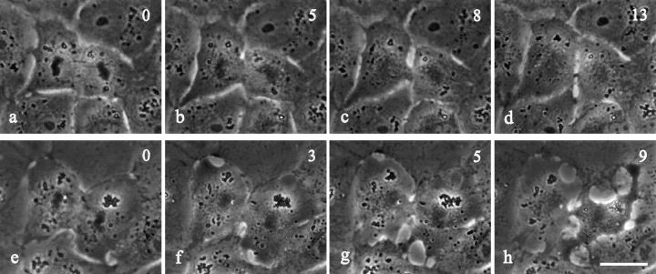
Figure 2.
Time lapse images of NRK cells microinjected with C3 transferase after the initiation of cytokinesis. Time in minutes after microinjection is shown in the upper right corner. Roughly half of the cells injected after the initiation of furrowing proceeded through cytokinesis without any apparent effects (compare a–d with Fig. 1, c–e). The remaining cells showed increased cortical activities outside the equatorial region (f–h). All the injected cells completed cytokinesis with a normal-looking furrow and midbody (d and h). Bar, 20 μm.

