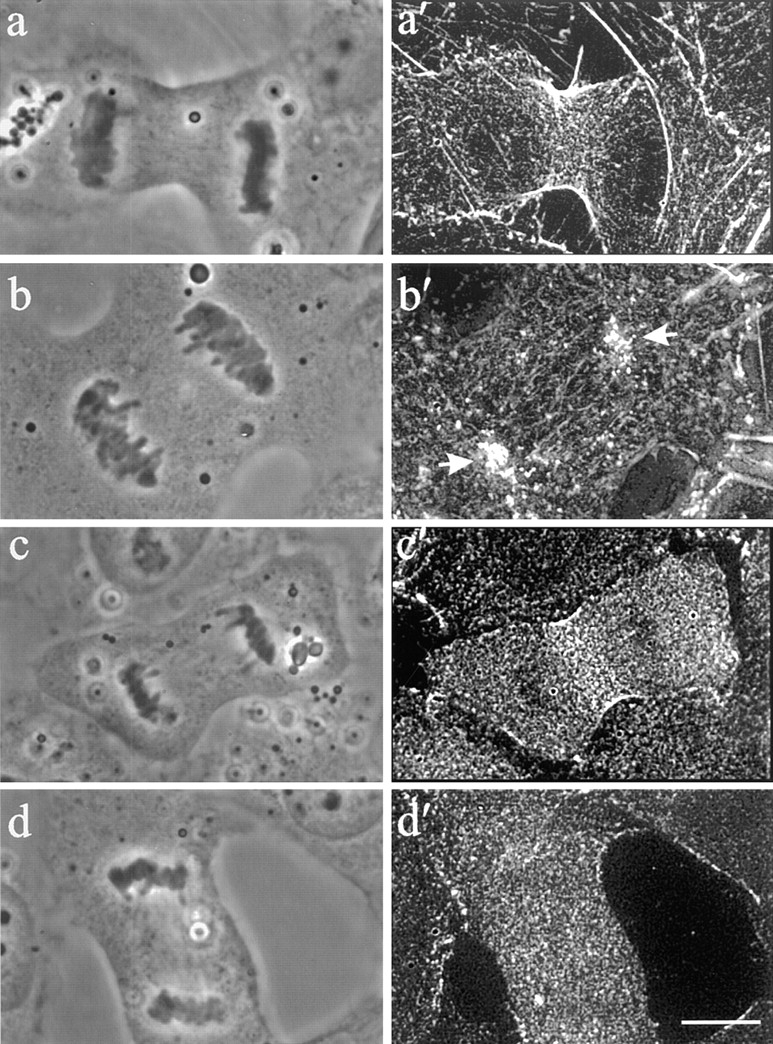
Figure 5.

The organization of F-actin and myosin II in control (a′ and c′) and Rho inhibited (b′ and d′) cells. Stacks of optical sections were deconvolved and used for reconstruction of the 90° view. Phalloidin staining of ingressions in C3-injected cells revealed clusters of cortical actin localized above separated chromosomes (b and b′, arrows). No equatorial accumulation of actin as found in control cells was visible (a and a′). Immunofluorescence of myosin II similarly demonstrated a lack of localization to the equatorial plane (d and d′) compared to control cells (c and c′). Myosin II remained diffuse throughout the cell and did not colocalize with cortical actin over the chromosomes. Bar, 10 μm.
