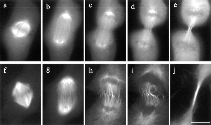
Figure 8.
Organization of microtubules in control and C3-injected mitotic NRK cells. In a control cell microinjected with rhodamine-labeled tubulin (a–e), kinetochore microtubules shortened, while midzone microtubules elongated. The spindle poles separated as the cell progressed from metaphase through anaphase (a–c). Prominent microtubule bundles were associated with the midbody during telophase (e). Microtubules in a C3- injected cell (f–j) underwent similar changes, except that midzone microtubules became distorted (compare c and d, with h and i). A long bundle of microtubules formed in the extended cleavage furrow (j). Bar, 10 μm.

