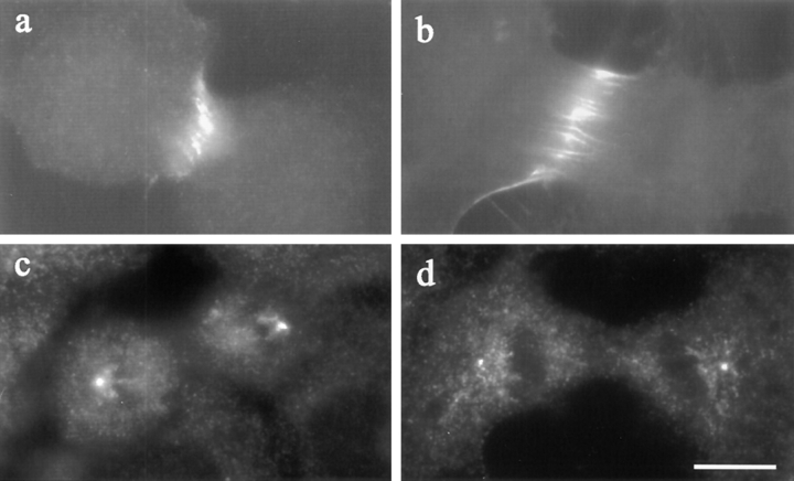
Figure 9.
The distribution of TD60 (a and b) and γ-tubulin (c and d) in Rho-inhibited NRK cells. Both control cells (a) and C3- injected cells (b) showed a localization of TD60 along the equator. Staining of γ-tubulin, used as a marker for centrosomes, showed similar localizations at the spindle poles in a C3-injected cell (d) and a control cell (c). Bar, 10 μm.

