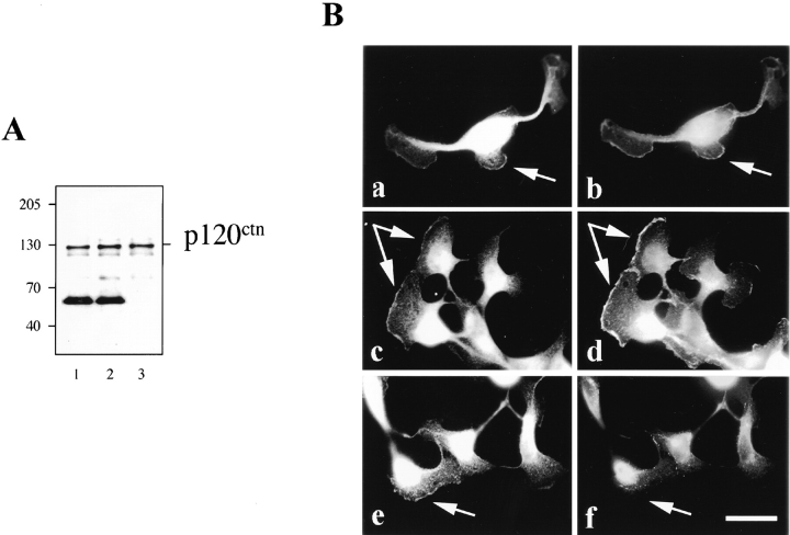
Figure 11.
Ectopic expression of δ-catenin does not lead to displacement of p120ctn from adhesive junction proteins. (A) Immunoblot showing p120ctn coimmunoprecipitation with E-cadherin in mock (1), and MF cells (2). (3) MDCK cell lysate. (B) Double immunofluorescent microscopy showing redistribution of junctional proteins in δ-catenin–transfected MDCK cells after HGF stimulation. (a, c, and e) Anti–δ-catenin. (b) Anti– β-catenin. (d) Anti-p120ctn. (f) Anti-desmoplakin. Note the colocalization of δ-catenin with β-catenin and p120ctn, but not desmoplakin, at the leading edges of cells treated with HGF. Arrows point to the lamellipodia formation. Bar, 5 μm.

