Figure 2.
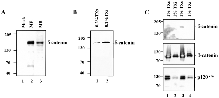
Expression of δ-catenin cDNA in transfected MDCK cells and endogenous δ-catenin in developing mouse brains. (A) Immunoblot showing δ-catenin expression in MDCK cells and developing mouse brains. (1) Mock-transfected MDCK cells; (2) MF (MDCK cells transfected with full-length δ-catenin cDNA); (3) MB (mouse brain lysate). (B) Immunoblot of transfected MDCK cells showing δ-catenin in the 0.2% Triton X-100 soluble (TXs, lane 1) and insoluble (Txi, lane 2) fractions. (C) Immunoblots of mock (1 and 2) and δ-catenin transfected (3 and 4) MDCK cells extracted in 1.0% Triton X-100. Soluble and insoluble fractions were blotted with the indicated antibodies. In all panels the molecular weight standard is indicated at the left.

