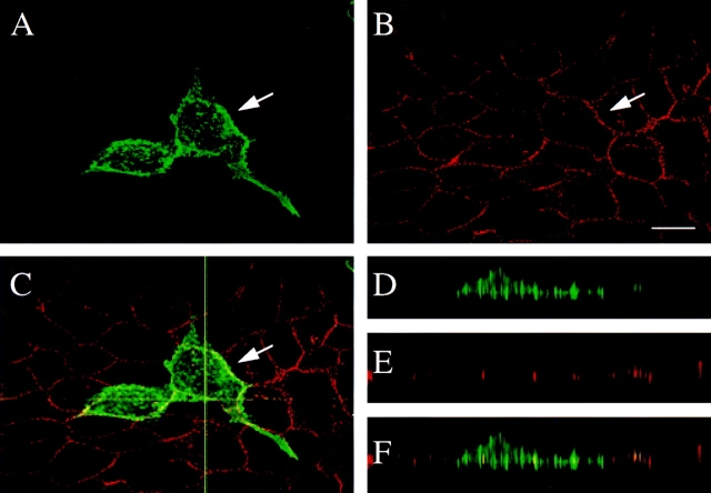
Figure 5.
Confocal immunofluorescence microscopy of MDCK cells transiently transfected with δ-catenin. The cells were double labeled with (A) δ-catenin antibodies and with (B) desmoplakin antibody. The arrow points to the transfected cell. (C) Merged fluorescent image showing minimal colocalization of δ-catenin and desmoplakin. The horizontal line indicates where the XZ plane was selected for D–F. (D–F) Respective XZ vertical sections of A–C. Bar, 15 μm.

