
Figure 1.
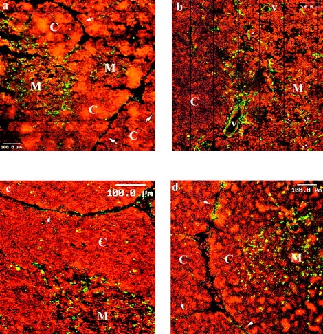
Immunofluorescence on frozen human thymus section was performed with anti-laminin 5 chain mAbs: the anti-α3 chain BM165 (a and b), the anti-β3 chain 6F12 (c); the anti-γ2 chain GB3 (d). mAb staining was revealed by FITC conjugated goat anti–mouse F(ab)′2 fragments and analysis was performed by laser scanning confocal microscopy. Variations of cellular density between cortex (C) and medulla (M), were revealed by cell nuclear staining with propidium iodide. Single arrows designate the subcapsular basal laminae. Double arrows show examples of laminin 5 positive epithelial cells. b is a view constructed by the juxtaposition of successive areas scanned at a larger magnification.