
Figure 2.
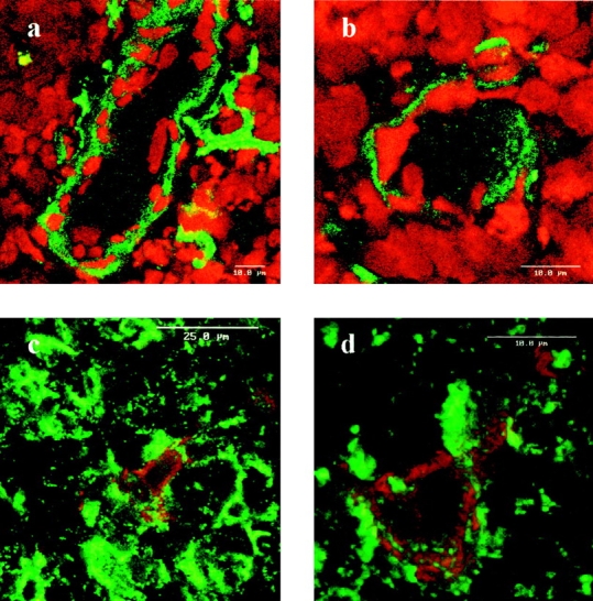
Frozen human thymus section stained with anti-laminin 5 α3 chain mAb BM165. (a and b) BM165 staining on vessels; staining was revealed by FITC conjugated goat anti–mouse F(ab)′2 fragments and analysis was performed by laser scanning confocal microscopy. Cell nuclei were stained with propidium iodide. (c and d) BM165 staining on stellate thymic epithelial cells; double staining obtained with mAb BM165 revealed by rabbit anti– mouse IgG1 F(ab)′2 fragments followed by PE conjugated goat anti–rabbit F(ab)′2 fragments, and the anti-keratin CK19 revealed by FITC conjugated goat anti–mouse IgM Ab. Controls made with secondary Abs gave no staining (not shown).