
Figure 2.
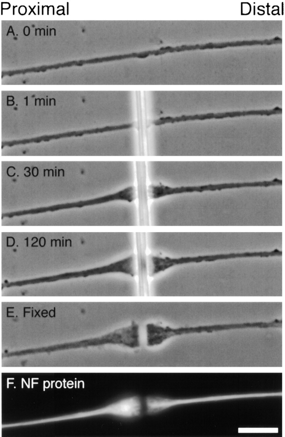
Morphological changes at the site of constriction. Phase contrast images of an axon immediately before constriction (A) and after constriction for 1, 30, and 120 min (B–D). The cell was fixed after constriction for 2 h and then the glass fiber was removed (E) and the cell was stained for NF-L by immunofluorescence microscopy (F). Bar, 5 μm.