
Figure 8.
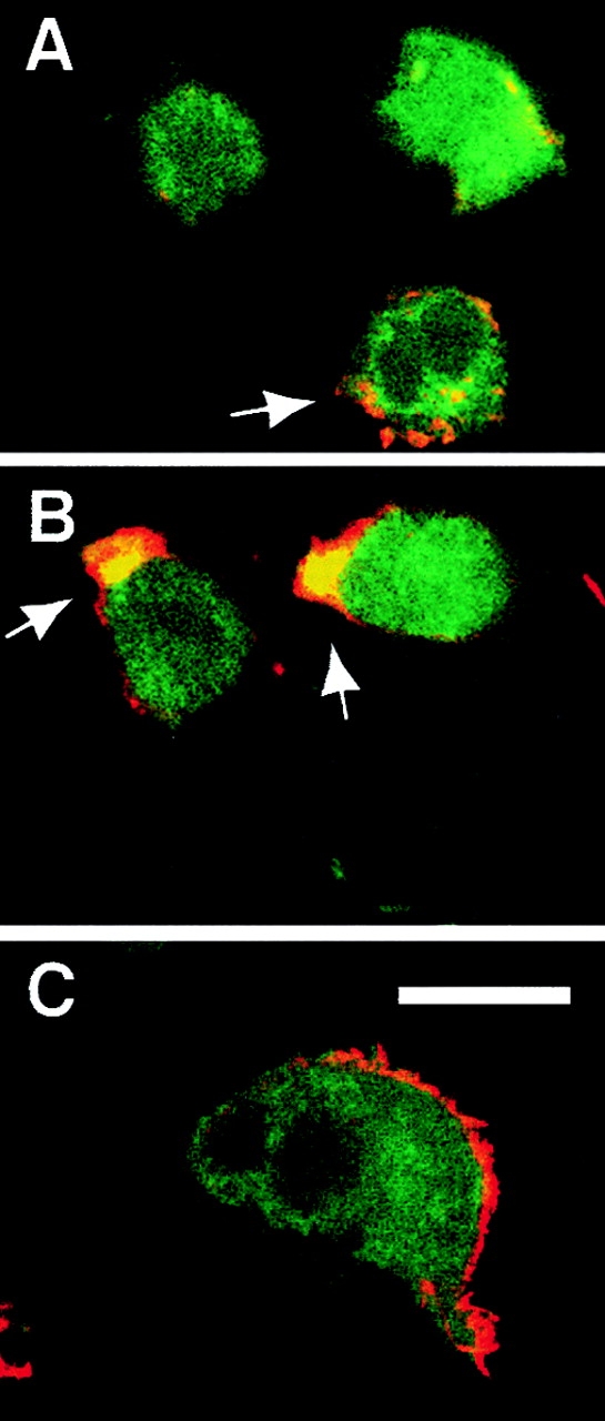
Confocal immunofluorescent microscopic examination of Syk localization during LCL attachment. LCL were allowed to attach for 10 (A and B) or 30 (C) min and subsequently fixed and stained for the presence of endogenous Syk with monoclonal 4D10 followed by secondary detection by FITC-conjugated goat anti– mouse (green), and costained with rhodamine-conjugated phalloidin to label F-actin (red). Significant colocalization of the signals (yellow) did not occur in nonadherent LCL (A, arrow) and was modest in confocal Z sections of adherent LCL viewed above the adhesive substrate (A) (Z plane = 11 μm above the substrate). Colocalization of signal was observed consistently in the podosomes (B, arrows) of actively attaching LCL at the cell–substratum interface (Z plane = 1 μm above the substrate), but not in fully spread cells at 30 min (C) (Z plane = 1 μm above the substrate). All Z sections are 1 μm. Bar, 10 μm.