
Figure 3.

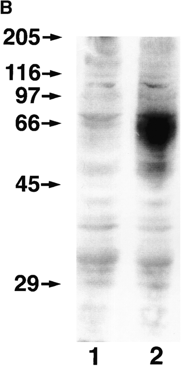
Cell surface expression of LYVE-1 receptor on transfected COS cells. In A, COS 1 cells were transiently transfected with either full-length LYVE-1 cDNA in the expression vector pRcCMV (a and b), or with a control empty pRcCMV vector (c and d) using DEAE dextran followed by surface immunofluorescent staining with rabbit polyclonal LYVE-1 antiserum (1:100 dilution) and FITC goat anti–rabbit IgG. In B, control and LYVE-1 transfected COS cells were electrophoresed on a 10% polyacrylamide SDS-PAGE gel, transferred to nitrocellulose, and Western blotted with LYVE-1 antiserum and peroxidase-conjugated goat anti–rabbit IgG (see Materials and Methods). Samples were control transfected COS (lane 1) and LYVE-1 transfected COS (lane 2). The positions and sizes in kilodaltons of the molecular mass calibration markers myosin (205 kD), β-galactosidase (116 kD), phosphorylase b (97 kD), BSA (66 kD), ovalbumin (45 kD), and carbonic anhydrase (29 kD) are indicated on the left.