
Figure 8.
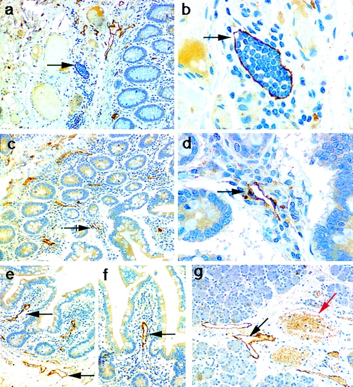
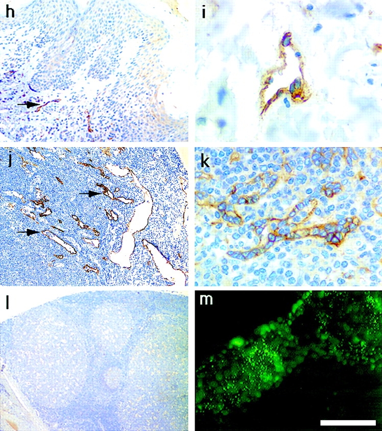
Localization of the LYVE-1 HA receptor in human tissues. Paraffin-embedded human tissue sections were stained with rabbit polyclonal LYVE-1 antiserum (1:100 dilution), followed by peroxidase-conjugated goat anti–rabbit IgG as described in Materials and Methods. Tissues shown are colon (a and b), small intestine (c–f), salivary gland (g), skin (h and i), lymph node (j), spleen (k), and tonsil (l). m shows cultured HUVEC stained with LYVE-1 and FITC-conjugated anti–rabbit Ig (see Materials and Methods). Bars, j and l, 500 μm; a, c, and e–h, 200 μm; b, d, and k, 50 μm; and i and m, 20 μm. Black arrows depict lymphatic vessels, both empty (d–g) and containing lymphocytes (b), and red arrows depict blood vessels clearly identified by their content of weakly stained erythrocytes (g and i).