
Figure 2.

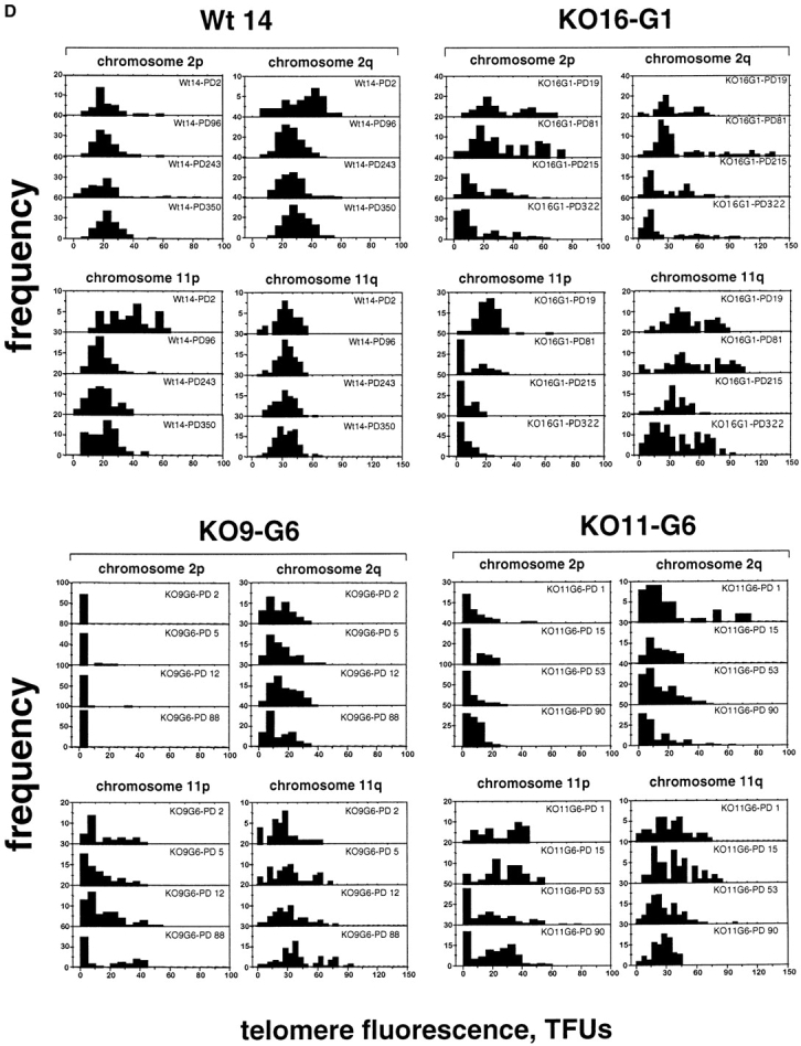
Telomere dynamics in wt and mTER−/− cell lines. (A–C) The telomere fluorescence, measured as TFU, of all telomeres (A), chromosome 2 telomeres (B), and chromosome 11 telomeres (C) in wt (Wt14) and mTER−/− cell lines at different PDs is shown. Black bars, fluorescence of q-telomeres; gray bars, fluorescence of p-telomeres. Despite the wide heterogeneity in individual telomere fluorescence intensity values, and due to the large number of data points used in the analysis, the error bars are not always visible in the graphs (i.e., A). (D) The distribution of telomere fluorescence intensity values of 2p-, 2q-, 11p-, and 11q-telomeres (black bars) in wt cell line (Wt14) and the indicated mTER−/− cell lines at increasing PDs.