
Figure 1.
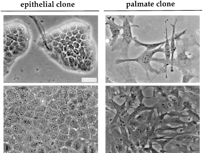
Morphology of clones derived from the MMH E14 line. Phase-contrast micrographs of the epithelial and the palmate clones at low (top) and high or intermediate (bottom) cell density, respectively. In the micrographs of the cultures at low density the differences in growth habit and cellular shape between the epithelial and the palmate clone are highlighted; those at higher density document that these differences are maintained. Bar, 40 μm.