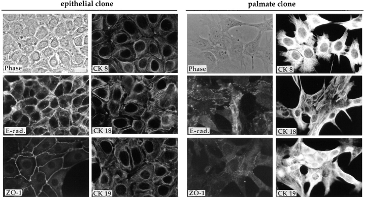
Figure 2.
Immunofluores- cence analysis of expression of epithelial and stage-specific markers of the hepatic cell lineage (CKs 8, 18, and 19). Phase-contrast micrographs and immunofluorescence staining of the E14 epithelial and palmate clones for the expression and localization of E-cadherin (E-cad.) and ZO-1, and of the three CKs 8, 18, and 19. (The cytokeratin antibodies were used on liver sections as a control where they showed the expected staining patterns: CK 8 and 18 on hepatocytes, and CK 19 primarily on bile ducts). For the epithelial clone the E-cad. staining and the phase-contrast image are of the same field; for the palmate clone the CK 8 field corresponds to the phase-contrast image. Bar, 20 μm.

