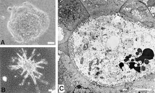
Figure 4.

Differences in the intrinsic morphogenic activity of the epithelial and the palmate clones are revealed by culture on the three-dimensional matrix, Matrigel. Photographs taken from 10-d cultures on a thick layer of Matrigel. (A) Phase-contrast micrograph of the spheroidal organization of the epithelial clone; (B) palmate cells, showing a spheroidal colony sending out tubules. (C) composite of four overlapping electron micrographs of a thin section across a tubule of palmate cells highlighting a duct-like structure with a well-defined lumen and circumscribed by well-polarized cells with junctional complexes and luminal membranes covered with microvilli. Bars: (B) 40 μm; (C) 5 μm.
